


- Welcome
- Shoulder instability
Shoulder instability
Shoulder
Introduction
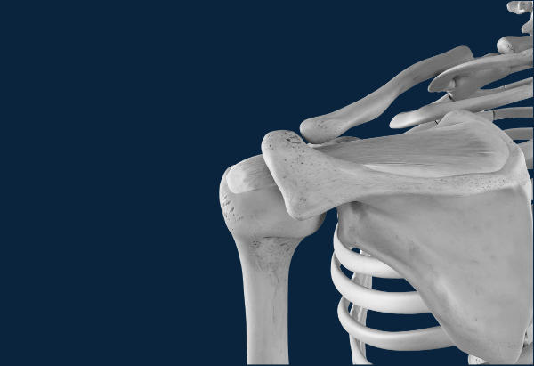
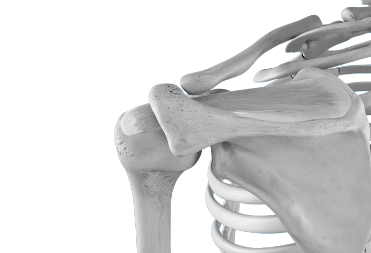
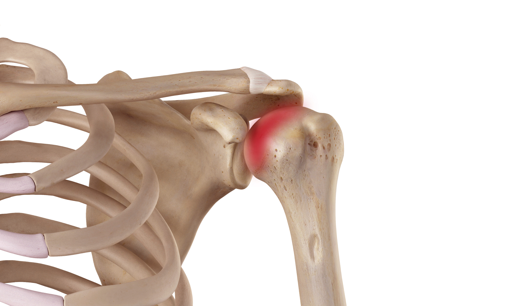
The shoulder is remarkable for its range of motion in all directions thanks to the joints that compose it:
- Sternoclavicular joint
- Acromioclavicular joint
- Glenohumeral joint
- Scapulothoracic joint (sliding of the shoulder blade on the thorax).
To animate the shoulder and at the same time stabilize it by pressing the articular surfaces against each other, there is a set of very effective small muscles grouped under the term "rotator cuff" because of their anatomical arrangement above the head of the humerus.
These rotator muscles of the shoulder are:
- the supraspinatus (Supraspinatus)
- the infraspinatus
- the subscapularis
- the small circle (Teres Minor).
The supraspinatus also plays a key role in raising the arm and allows the head of the humerus to slide under the bony roof of the shoulder called the "acromion," which is part of the scapula.
Signs & symptoms
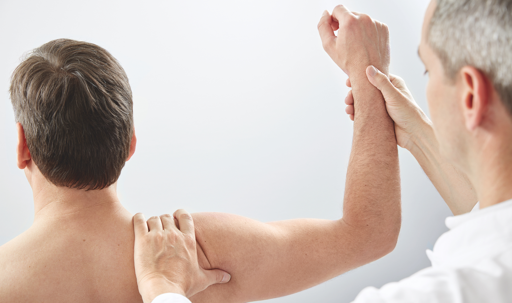
Symptoms vary depending on the type of instability and the direction of dislocation or subluxation of the humeral head. Certain factors are predisposing, and instability can sometimes be multidirectional.
In the acute stage, a traumatic dislocation is accompanied by significant pain and an inability to use the upper limb. This situation requires a medical evaluation and reduction of the dislocation as quickly as possible. Once the shoulder is reduced, immobilization is standard practice, followed by appropriate physiotherapy. Nevertheless, and particularly in young patients, the risk of instability and re-dislocation is high. One of the symptoms frequently encountered is fear of dislocation: this instability appears when the arm is raised if it is an anterior dislocation, the most common type.
Shoulder instability is a symptom that feels like the joint might dislocate during certain movements. Because the shoulder is a suspended and poorly congruent joint (with its structures not fitting together properly), instability develops more easily than in other joints.
What is this?
We distinguish between so-called traumatic and post-dislocation instability, and so-called atraumatic instability which is found in hypermobile subjects.
Traumatic instability is the result of an accident, usually a shoulder dislocation. It is generally present on only one side.
The risk of recurrence is inversely proportional to age:
- 90 to 95% risk of recurrence for an age under 20 years.
- 60% of recurrence for patients aged 20 to 40 years.
- 10% of recurrence only after 40 years.
The frequency of lesions observed on the rotator cuff tendons is proportional to age, and therefore one must be wary of such lesions in patients over 40 years of age.
Surgical treatment is particularly recommended in young patients where the risk of recurrent dislocation is high and where functional impairment is significant given the activities performed.
Instability of non-traumatic origin is not linked to an accidental cause and frequently manifests itself in everyday activities such as brushing hair or sleeping at night.
Both shoulders are usually affected and significant flexibility, known as hyperlaxity, is also observed in other joints, reflecting a loose texture of the joint capsule.
Treatment is primarily conservative, combining physiotherapy and joint-protective exercises. Surgical capsular repair may be considered in certain specific cases.
Each case must be analyzed individually and receive the most appropriate treatment, taking into account post-traumatic or predisposing factors.
Risk factors
The main risk factors in cases of traumatic dislocation are:
- Sports activities with a risk of falls.
- Young and active patients.
- Ball sports using the "army" position of the arm (volleyball for example) can cause repeated trauma ultimately leading to instability.
Non-traumatic instability is similar to hyperlaxity aggravated by:
- Impaired joint control.
- Repeated micro-lesions, the dominant arm in case of prior instability, neurological or psychological factors.
Screening & Diagnosis
In case of shoulder instability or dislocation, particular attention should be paid to:
- the manner in which the dislocation occurred
- the position that predisposes one to a feeling of instability
- the frequency of the disorders and their occurrence
A radiological assessment including at least two views must be carried out in order to determine the direction of the dislocation and the presence of any joint fracture or tendon avulsion.
Magnetic resonance imaging is required on a case-by-case basis depending on the indication for surgical treatment or the risk of associated injuries, particularly affecting the rotator cuff.
Treatments
Self-treatment
In case of shoulder instability or dislocation, particular attention should be paid to:
- the manner in which the dislocation occurred
- the position that predisposes one to a feeling of instability
- the frequency of the disorders and their occurrence
A radiological assessment including at least two views must be carried out in order to determine the direction of the dislocation and the presence of any joint fracture or tendon avulsion.
Magnetic resonance imaging is required on a case-by-case basis depending on the indication for surgical treatment or the risk of associated injuries, particularly affecting the rotator cuff.
Medical treatments
Once the diagnosis of dislocation has been made and demonstrated radiologically, the doctor reduces the joint into the correct position using appropriate analgesia or even anesthesia if necessary.
If this is a first-time dislocation, post-traumatic treatment involves immobilizing the shoulder in a body bandage for three weeks, immediately beginning physiotherapy with muscle strengthening and joint stabilization exercises. These exercises will then be gradually intensified, avoiding movements that could put stress on the joint capsule where it needs to heal. The patient will gradually regain mobility, and the need for surgical stabilization will be discussed based on residual instability and functional limitations.
Our favorite methods
Each case requires a personalized assessment in order to make an accurate assessment of the lesions and to decide on the choice of the most appropriate treatment based on the anatomical damage, activities and personal request.
Anterior shoulder instability is by far the most common. Orthopedic treatment primarily involves stabilizing the scapula, strengthening the internal rotators, and protecting the joint during high-risk activities.
The most common surgical treatment involves restoring the anatomy by retensioning and reattaching the capsule to the anterior edge of the glenoid cavity of the scapula, the most frequent site of shoulder stabilizing lesions. This procedure, generally called a Bankart repair, is performed either open or arthroscopically. Arthroscopic treatment is more frequently used and is recommended because it facilitates postoperative rehabilitation. Open surgery is preferred in cases of chronic lesions or associated joint and bone damage.
Surgery
Preparation for surgery
Your general practitioner
We are available to inform your primary care physician about the proposed treatment so that we can provide you with the best possible care, prevent risk factors, and ensure optimal postoperative follow-up. Once the surgical treatment is scheduled, your primary care physician may be asked to complete sections of a form that will be sent to the anesthesiologist. This request is standard practice for major procedures.
An appointment with the anesthesiologist may be deemed useful or essential depending on the type of procedure and any existing risk factors. This appointment takes place before or upon your admission to the clinic, or even on the day of the operation itself, for example. The purpose of this consultation is to provide you with all the necessary information regarding the type of anesthesia, which will be decided upon in consultation with you. The discussion will cover not only the anesthesia itself but also methods for managing pain during the postoperative period.
Depending on the surgical schedule, you will be admitted the day before your procedure or on the same day, 3 hours before the operation, on an empty stomach.
Recovery
The first postoperative day:
Treatment focuses primarily on pain relief and restoring free movement to the cervical and thoracic spine. Massage and mobilization of the scapula are also performed, and depending on the pain level, the patient is encouraged to roll their shoulders forward and backward.
The second postoperative day:
Pain relief treatment through massage and cervico-dorsal mobilization. Pendulum mobilization of the elbow and shoulder. Shoulder rolling movement and initiation of scapular stabilization exercises.
Usually, on the third postoperative day, The patient can return home with an exercise program focused on isometric shoulder strengthening (exercises that can be done without moving the joint), shoulder mobilization using a pendulum motion, shoulder rolling movements, scapular stabilization exercises, and the start of the program in the pool.
Possible risks
The infection
Infection is always a serious complication in orthopedic surgery, but fortunately very rare (less than 0.51 TP3T). Specific measures are taken to minimize these risks, including: systematic pre- and immediate post-operative antibiotic therapy, operating rooms equipped with high-flow laminar airflow, a highly trained and experienced orthopedic surgical team, optimal skin preparation, and a careful assessment of your health.
If you experience fever or local redness after your procedure once you have returned home, you must call your surgeon immediately.
Limitation of shoulder mobility
Due to inflammation and scar tissue formation following surgery, shoulder mobility may be temporarily reduced. A physiotherapy program is essential to regain full shoulder mobility, strength, and stability. Because this procedure involves anatomical reconstruction of the joint capsule, there is a risk of recurrent dislocation or instability in the event of subsequent trauma or high-risk activities.
Frequently Asked Questions
What type of anesthesia is offered for this type of surgery?
How long should I wear a scarf or vest?
When will I be able to start driving again?
Summary
1006 Lausanne
+41215103348
Learn more in videos
Medicol Videos
Contact one of our specialists on the subject
Ouchy Orthopedic Center
Specialized medical consultations in orthopedics and traumatology in the heart of Lausanne