


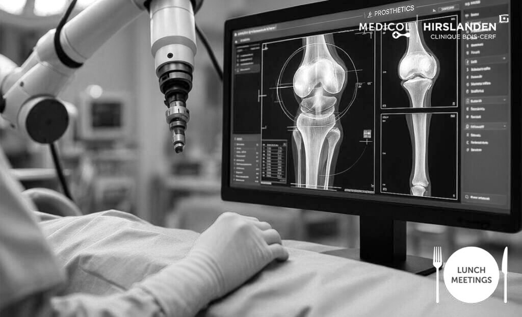

- Welcome
- Functional hallux limitus
Functional hallux limitus
Introduction
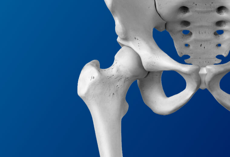
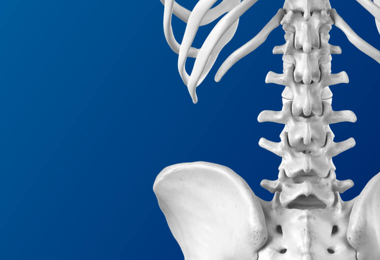
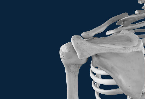
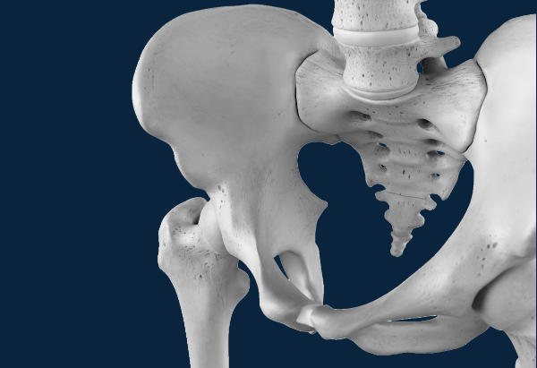
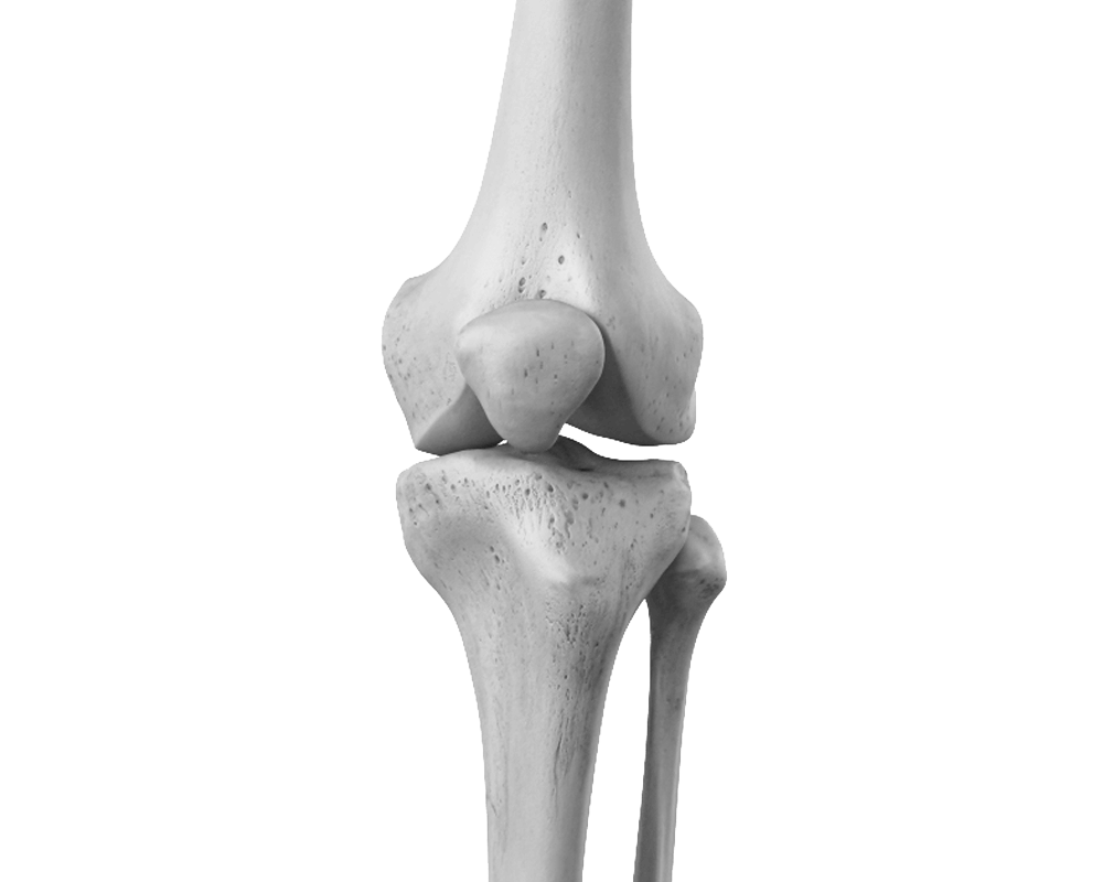
Anatomy

Anatomy
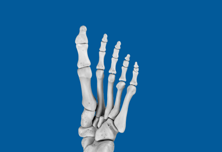
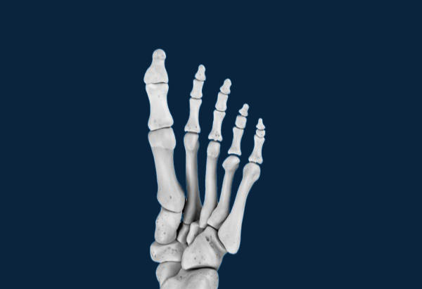
The flexor hallucis longus muscle originates on the deep posterior aspect of the leg and inserts distally on the distal phalanx of the big toe. Its action is pronatory in the foot, propulsive during walking, and, as its name suggests, flexes the big toe. The inability to glide at the retrotalar pulley results in what is called a tenodesis effect, a taut rope-like effect under the sole of the foot that occurs when the ankle is dorsiflexed. This "rope" effect causes automatic plantar flexion of the first toe.
Signs & symptoms
Deformation:
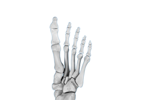
Locally, on the big toe, hyperkeratosis or corns are frequently observed on the inner edge of the distal phalanx and on the inner edge of the base of the head of the first metatarsal. These corns indicate a syndrome of increased pressure and local friction related to weight transfer. A claw deformity of the second toe and sometimes hyperextension of the interphalangeal joint of the big toe are frequently observed in association.
Locally, symptoms may be related to excessive pressure but may also present as painful tension under the sole of the foot located on the tendinous path of the long flexor hallucis, pain under the 1st metatarsal head corresponding to a tendency to subluxation of the sesamoids, pain in the posterior part of the hindfoot opposite the tendon conflict under the retro-talar pulley.
Symptoms may also be present at a distance in the form of patellar syndrome, pes anserine tendinitis or iliotibial band syndrome, painful contracture of the piriformis muscle or lumbopelvic imbalance.
What is this?
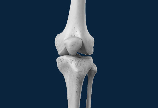
Functional Hallux Limitus, or Functional Hallux Rigidus, is defined during gait by the inability to extend the first metatarsophalangeal (MTP1) joint during the gait cycle. During the propulsive phase, when the ankle is in dorsiflexion, the MTP1 joint remains rigid and cannot dorsiflex. This biomechanical condition results from a tenodesis of the flexor hallucis longus tendon in the hindfoot, preventing it from gliding freely.
From a biomechanical perspective, this dysfunction has significant repercussions, particularly on foot stability.
Functional hallux limitus, or functional hallux rigidus, is a relatively common but little-known condition because it is often asymptomatic locally and difficult to diagnose. Tests to detect tendon blockage of the flexor hallucis longus are indeed not widely used, nor are tests to assess the mobility of the subtalar joint.
The functional disorders caused by hallux rigidus are not limited to the foot but also affect the knee, hip, and lumbopelvic region. The disruption of the normal gait cycle leads to joint overload and compensatory mechanisms elsewhere in the body.
Risk factors
The presence of functional hallux limitus or functional hallux rigidus is primarily linked to muscle hypertrophy or a very distal musculotendinous junction of the flexor hallucis longus, or to an inflammatory condition such as tenosynovitis. Less frequently, tendon nodules or instability of the subtalar joint are observed.
The patients' medical history frequently reveals repeated ankle sprains, ankle immobilization for a leg fracture or other lower limb condition, a tendency to walk on tiptoe during childhood, participation in classical dance or figure skating, and forefoot deformities in the family.
Screening & Diagnosis
Functional Hallux Limitus or Functional Hallux Rigidus can be diagnosed clinically using a specific test called the "flexor hallucis longus stretch test." During a podiatric assessment, the footprint image is pathognomonic. Finally, gait analysis on a treadmill reveals the inability to dorsiflex the MTP1 joint during ankle dorsiflexion.
The flexor hallucis longus stretch test is performed in three phases: first, the patient lies on their back, placing their ankle in plantar flexion, and the free movement of the first metatarsophalangeal (MTP1) joint is verified. Second, the ankle is placed in maximum dorsiflexion by applying pressure under the ball of the foot at the metatarsals. Third, an attempt is made to dorsiflex the first metatarsophalangeal (MTP) joint by pushing the toe backward. If the toe can be pushed backward, the test is negative. If, however, it is impossible to push the toe backward, the test is positive and demonstrates tendon blockage in the hindfoot.
Dynamic foot tracing during a gait or running examination reveals a lack of support under the head of the first metatarsal and excessive pressure under the ball of the big toe. A static foot assessment shows a shift in the projection of the center of gravity towards the rear and outside of the foot.
Treatments
Self-treatment
Certain stretching exercises are helpful for improving the gliding of the flexor hallucis longus tendon once the subtalar joint has been freed by manipulation. However, performing the joint manipulations oneself is difficult.
Initially, physiotherapy or osteopathy treatment, or a combination of both, is necessary.
Medical treatments
The treatment aims to release the tenodesis (or cord effect) of the flexor hallucis longus tendon as it passes under the retrotalar pulley. This release can be achieved through orthopedic treatment by manipulating the subtalar joint. A specific maneuver has been described to release the tendon blockage, which we have called the "vacuum cleaner cord" maneuver, by analogy to a vacuum cleaner cord that, when bent, gets caught under a table leg and remains stuck there despite all the traction applied. To release the tendon, as with releasing the vacuum cleaner cord, it is necessary to change the axis of traction. At the level of the foot, traction is applied along the axis of the calcaneus, using internal and external decoupling movements, gradually freeing the subtalar joint. As this is done, the tendon is released, and the stretch test becomes negative. Tendon gliding can then be trained by performing specific stretching exercises. Orthopedic treatment based on this maneuver and the repetition of stretching exercises over a sufficient period of time generally gives good results.
If this fails, surgical treatment is performed in the form of a surgical release of the retrotalar pulley of the flexor hallucis longus. Initially, this technique was performed via open surgery through a 3 cm incision in the medial retromalleolar region. Now, it is performed endoscopically using two small incisions located on either side of the Achilles tendon. After arthroscopy of the subtalar joint, the retrotalar pulley of the flexor hallucis longus is located and incised at the point where it meets the talus. Free tendon gliding and functional recovery of subtalar joint mobility are then confirmed, and the two arthroscopic incisions are closed with a single suture.
Our favorite methods
Initially, patients are referred to physiotherapy where they are offered a standardized treatment plan including hindfoot mobilization, stretching exercises, and helpful daily lifestyle precautions. In some cases, this treatment may be supplemented by a prescription for orthotics, depending on the sports activities they participate in.
If these measures fail, endoscopic surgery is recommended. The arthroscope is inserted through a posterolateral approach on the lateral aspect of the Achilles tendon. This approach allows observation of the subtalar joint and identification of the retrotalar pulley of the flexor hallucis longus. Instruments are then inserted through an incision on the medial aspect of the Achilles tendon to cut the pulley at the point where it contacts the talus, until complete tendon gliding is achieved and free movement of the subtalar joint is restored. After final hemostasis control, the arthroscopic incisions are closed with a single suture.

Surgery
Preparation for surgery
Your family doctor's involvement
We will be in constant communication with your family doctor to discuss and proceed with the best treatment option for you. Once surgery has been indicated, your doctor will arrange the necessary tests to complete the anesthesiologist's pre-operative assessment.
You will receive a summons letter from the Bois-Cerf Clinic with information regarding:
Pre-anesthesia consultation
Before surgery, you will be seen by one of our anesthesiologists who will ask you about your medical history before conducting a clinical examination. You will then be able to discuss the different methods available and choose the most suitable one for your specific case (general anesthesia, epidural anesthesia, spinal anesthesia, regional venous anesthesia, nerve blocks, combined anesthesia).
– What day and what time should I arrive at the reception of the Bois-Cerf Clinic? It could be the day before or the day of the procedure.
Surgery
What you need to bring with you:
- Your personal belongings
- Reading material if you enjoy reading
- Music player
- Your personal medications (if you are receiving treatment at home)
- Your optimism and energy... we'll take care of the rest.
Post-operative pain management
PAIN AFTER THE OPERATION IS UNACCEPTABLE FOR US.
The night before your surgery and again in the hour before your anesthesia, you will receive premedication (a tablet to help you relax). The anesthetic strategy will be specifically tailored to your health, your preferences, and the type of surgery, with the aim of ensuring you experience no pain upon waking and during your rehabilitation.
Bed rest and ice will be helpful during the first few days. Our team will carefully assess the presence and intensity of your pain with you 24/7 and adjust your treatment accordingly.
Recovery
The day after the procedure, the patient is able to walk without crutches or supports. Initially, walking is done with small steps, then normally after a few days. A physiotherapy program is prescribed to restore free tendon movement and rebalance the entire lower limb by correcting compensatory mechanisms.
Possible risks
The infection
Infection is always a serious complication in orthopedic surgery, but fortunately very rare (less than 0.51 TP3T). Specific measures are taken to minimize these risks, including: systematic pre- and immediate post-operative antibiotic therapy, operating rooms equipped with high-flow laminar airflow, a highly trained and experienced orthopedic surgical team, optimal skin preparation, and a careful assessment of your health.
If you experience fever or local redness after your procedure once you have returned home, you must call your surgeon immediately.
Bleeding
The risk of bleeding exists but is relatively low in this case given that it is an arthroscopic approach.
Subtalar instability
The subtalar joint consists of an anterior and a posterior portion separated by the sinus tarsi. If the ligaments within this space are intact, the risk of instability is low. However, if the ligament is torn and there is constitutional hyperlaxity, endoscopic tenolysis of the flexor hallucis longus (FHL) can, in some cases, lead to further laxity of the subtalar joint.
Frequently Asked Questions
How long does the operation take?
At what point should the wires be removed?
When will I be able to walk with full weight-bearing?
Summary
1006 Lausanne
+41215103348
Learn more in videos
Medicol Videos

9:44

42:09

43:20
Contact one of our specialists on the subject
Ouchy Orthopedic Center
Specialized medical consultations in orthopedics and traumatology in the heart of Lausanne